Hong Chen1 ![]() ,
Xue-mei Wan2,
Xue-lei Zhou3
,
Xue-mei Wan2,
Xue-lei Zhou3
For correspondence:- Hong Chen Email: woshichenh2007@163.com Tel:+862365311341
Received: 13 June 2016 Accepted: 9 October 2016 Published: 29 November 2016
Citation: Chen H, Wan X, Zhou X. Anti-thrombotic and anti-tumor effect of water extract of caulis of Sargentodoxa cuneata (Oliv) Rehd et Wils (Lardizabalaceae) in animal models. Trop J Pharm Res 2016; 15(11):2391-2397 doi: 10.4314/tjpr.v15i11.13
© 2016 The authors.
This is an Open Access article that uses a funding model which does not charge readers or their institutions for access and distributed under the terms of the Creative Commons Attribution License (http://creativecommons.org/licenses/by/4.0) and the Budapest Open Access Initiative (http://www.budapestopenaccessinitiative.org/read), which permit unrestricted use, distribution, and reproduction in any medium, provided the original work is properly credited..
Purpose: To investigate the anti-thrombosis and anti-tumor effect of the water extract of the caulis of Sargentodoxa cuneata (Oliv.) Rehd. et Wils. (WCSW) in rat and mouse models.
Methods: WCSW extract was prepared and the main constituents were determined by high pressure liquid chromatography (HPLC). The acute toxicity of the extract was determined in mice. Platelet aggregation in rat platelet-rich plasma (PRP) was examined to evaluate the effect of the extract on platelet function. Thereafter, the cytotoxic activity of WCSW on HL60, A549, S180 and H22 cells was determined by 3-(4,5-Dimethylthiazol-2-yl)-2,5-diphenyltetrazolium bromide (MTT) assay. In vivo anti-tumor effect of WCSW was further evaluated on H22 cells transplanted in mice, while the ex
Results: Protocatechuic acid, rhodiola glucoside and chlorogenic acid were identified as the main constituents of WCSW. Platelet aggregation was significantly inhibited by treatment with the extract at concentrations of 1, 5 and 10 mg/mL. WCSW also showed significant inhibitory effect on HL60, A549, S180 and H22 cells in vitro with half maximal inhibitory concentration (IC50 value of 321.9, 285.0, 130.3 and 76.1 μg/mL, respectively. Furthermore, WCSW exhibited obvious anti-tumor effect on H22 transplanted tumor in vivo. After treatment with WCSW, caspase-3, caspase-9 and Bax were significantly (p < 0.05) up-regulated, whereas Bcl-2 was significantly (p < 0.05) down-regulated in the tumor tissues.
Conclusion: WCSW possesses significant antithrombosis and anti-tumor effect, and therefore, has the potentials to be developed into effective drugs for clinical treatment of cancer and thrombosis diseases.
Introduction
Thrombus formation is a pivotal event involved in the pathogenesis of cardiovascular disease, and platelets are critical in all phases of thrombus formation [1]. Antithrombotic drugs, such as adenosine diphosphate (ADP) receptor blockers, acetylsalicylic acid (aspirin), and glycoprotein (GP) IIb/IIIa antagonists are used to prevent cardiovascular disease [2]. However, these drugs can have systemic hemorrhagic side effects [3,4]. Cancer is a very serious medical condition worldwide. The current medical treatments for cancer including chemotherapy and radiotherapy, often have serious side effects [5,6]. Therefore, it is very important to develop much safer antithrombotic and anti-tumor agents with fewer side effects.
Sargentodoxa cuneata (Oliv.) Rehd. et Wils. (SRW), belonging to Lardizabalaceae, is a common Chinese medicinal plant which is distributed in South, East, Central and Southwest China [7]. The caulis of SRW is used in China for treating acute appendicitis, abdominal pain, rheumatic arthritis, trauma, amenorrhea and dysmenorrhea [8]. Several kinds of compounds such as lignans, triterpenes, anthraquinones, phenolic acids, and phenylpropanoids have been previously isolated from SRW [7,9]. However, to the best of our knowledge, only a few researches reported the pharmacological activities of SRW. The present research was aimed to investigate the antithrombosis and anti-tumor effect of water extract of caulis of SRW (WCSW), which may encourage the development and application of this drug.
Methods
Chemicals and reagents
Adenosine 5’-diphosphate (ADP), collagen, acetylsalicylic acid (AA) and 3-(4,5-Dimethylthiazol-2-yl)-2,5-diphenyltetrazolium bromide (MTT) were purchased from Sigma (St. Louis, MO, USA). Protocatechuic acid, Rhodiola glucoside, and Chlorogenic acid standards were purchased from National Institute for Food and Drug Control (Beijing, China). 5-Fluorouracil (5-FU) was obtained from Sinopharm Chemical Reagent Co., Ltd. (Shanghai, China). Caspase-3, caspase-9, Bcl-2, Bax, and β-actin antibodies were purchased from Beyotime Institute of Biotechnology (Nanjin, China). All the other chemicals used were of analytical grade.
Preparation of WCSW
Dried crude caulis of SRW was purchased from Chengdu Hehuachi Chinese Herbal Medicine Market (Chengdu, China) in August, 2015, and identified by the Oncology Department of Traditional Chinese Medicine (Chongqing Cancer Research Institute,Chongqing, China). A voucher specimen (no. HM2015-0807) was deposited in the laboratory herbarium of Chongqing Cancer Research Institute. The caulis of SRW was powdered and refluxed two times for 2 h each time with distilled water. The filtrates were concentrated under reduced pressure at 40 °C and the final yield of the extract (WCSW) was about 12.7 % (w/w).
Determination of major constituents of WCSW
An HPLC system consisting of Waters 2695 HPLC and UV detector (Waters, USA) was used to analyze the constituents in WCSW. The components of WCSW were isolated on a Diamonsil C18 (250 × 4.6 mm, with 5 μm particle size) column (Dikma, Beijing). The column temperature was controlled at 30 °C and the UV detector was set at 280 nm. The gradient elution system was composed of acetonitrile as solvent A and 0.05 % solution of phosphoric acid in water as solvent B, and was set as follows: 0-10 min, 95 % B; 10 - 35 min, 95 % - 84% B; 35 - 50 min, 84 % - 75 % B; 50 - 60 min, 75 % - 55 % B. The flow rate was 1.0 mL/min.
Animals
ICR mice (20 ± 2 g) and Sprague-Dawley (SD) rats (210 ± 10 g) were purchased from Dashuo experimental animal Co. Ltd. (Chengdu, China). Animals were maintained at 23 ± 2 °C and 55 ± 5 % humidity under a 12-h light/12-h dark cycle and free access to food and water. All the animal treatments were strictly in accordance with Directive 2010/63/EU on the Handling of Animals Used for Scientific Purposes [10]. The experimental procedures were approved by the Animal Care and Use Committee of Chongqing Cancer Research Institute (approval no. 201509-N-12).
Cell culture
The cell lines of A549, HL60, S180 and H22 were cultured in RPMI medium supplemented with 10 % fetal bovine serum and antibiotics (100 U/mL penicillin and 100 μg/mL streptomycin), and incubated at 37 °C in the presence of 5 % CO2.
Acute toxicity test
Acute toxicity of WCSW on mice was carried out according to a previous report [11]. Mice were divided into six groups (n=10) and fasted for 12 h prior to the experiment with free access to water. The animals were administered orally with a single dose of WCSW (10, 100, 500, 1000 and 2000 mg/kg), or distilled water for the control group. After that, the mice were observed for clinical signs and mortality at 0.5, 1, 2, 4, and 6 h after dosing and twice a day thereafter for 14 days.
Assessment of platelet aggregation
Platelet aggregation was determined using the turbidimetric method reported by Xin et al [12]. SD rats were lightly anesthetized with diethyl ether. Blood was drawn from the abdominal aorta and collected into blood collection tube containing 3.8 % sodium citrate (1:9, v/v). Platelet-rich plasma (PRP) was prepared by centrifuged at 800 rpm for 10 min. Platelet-poor plasma (PPP) was obtained by further centrifuged at 3000 rpm for 10 min. The PRP was diluted with PPP to make platelet concentration of 5 ×10⁸ /mL.
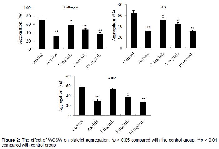
For the aggregation experiment, the diluted PRP was incubated at 37 °C for 5 min with different concentrations of WCSW (1, 5 and 10 mg/mL). Subsequently, arachidonic acid (0.5 mmol/L), ADP (5 μmol/L), or collagen (3.2 μg/mL) was added to induce platelet aggregation. Aspirin (25 μg/mL) was used as positive control. The changes in light transmission were recorded for 5 min.
MTT assay
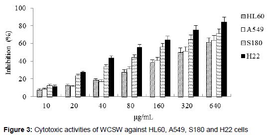
Cells were seeded into 96-well plates at a concentration of 1 × 10⁵/ 0.1 mL in the presence of WCSW (10 - 640 μg/mL) for 48 h. The growth inhibitory effect of the cells was determined by MTT assay. Briefly, each well was added 20 μL MTT (5 mg/mL) and incubated for another 4 h. Then the culture medium was removed, and 150 μL DMSO was added to each well. Absorbance was detected by a microplate reader at 570 nm. Inhibition (I) of cell proliferation was calculated by Eq 1.
I (%) = {(Ac- At)/Ac}100 ……………………. (1)
where Ac and At are the absorbance of control and treatment samples respectively.
In vivo antitumor experiment
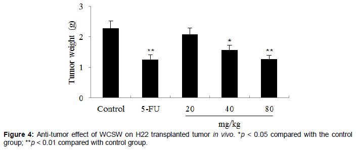
H22 tumor cell suspension (2 × 10⁶ cells/mouse, 0.2 mL) was subcutaneously injected into the right armpits of ICR mice to establish the animal model [13]. The mice were randomly divided into five groups (n = 10): the model control group, the positive group and three WCSW-treated groups (20, 40 and 80 mg/kg) by intragastric administration (ig). Control group was treated with normal saline {20 mL/kg, intraperitoneally (ip)} while the positive group was treated with 5-FU (20 mg/kg, ip). All the mice were sacrificed after treatment for 14 days, and the whole bodies and the segregated tumor of the mice were weighed immediately.
Western blot analysis
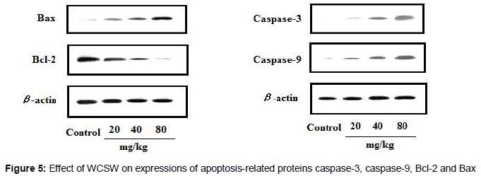
Total proteins were extracted from the tumor tissues. Protein samples (50 μg) were separated using sodium dodecyl sulfate polyacrylamide gel electrophoresis (SDS/PAGE). Thereafter, the separated proteins were transferred to a polyvinylidene difluoride (PVDF) membrane and probed with different primary antibodies targeting caspase-3, caspase-9, Bax, or Bcl-2 overnight at 4 °C. After washing with Tris buffered saline three times, the membrane was incubated with secondary antibodies. The proteins were detected using chemiluminescence peroxidase reagents. Antibody directed against β-actin was used to measure protein loading.
Statistical analysis
Data are expressed as mean ± standard deviation (SD). One-way ANOVA followed by t-test was performed on the data using SPSS software (SPSS for Windows 18.0, SPSS Inc, USA) to determine differences between the treated and control groups. P < 0.05 was considered statistically significant.
Results
Main constituents of WCSW
In order to detect the constituents in WCSW, HPLC was performed in the present study. As shown in , WCSW was well separated under the experimental conditions. Three constituents including protocatechuic acid, rhodiola glucoside and chlorogenic acid were identified by comparing their retention times and UV spectra with the corresponding reference standards.
Acute toxicity
Acute toxicity of WCSW was carried out in mice by oral administration. As a result, there was no death and significant modification in the general behavior of the animals after 14 days at all the administered doses of WCSW. Hence, WCSW was apparently safe when administered orally to mice under the studied doses.
Effect of WCSW on platelet aggregation in vitro
The effect on ADP, AA and collagen induced platelet aggregation in rat PRP were examined to evaluate the effect of WCSW on platelet function. Platelet aggregations induced by collagen and AA were significantly inhibited by treating with WCSW at the concentrations of 1, 5, and 10 mg/mL (p < 0.05, p < 0.05 and p < 0.01, respectively) in a concentration-dependent manner (). Furthermore, the inhibitory effect of WCSW on the platelet aggregations induced by ADP was also significant at the concentrations of 5 and 10 mg/mL (p < 0.05 and p < 0.01, respectively).
Cytotoxic activity of WCSW in vitro
The proliferation of HL60, A549, S180 and H22 cells was determined to investigate the cytotoxic activities of WCSW and the cell viability was determined by the MTT assay. The results in indicate WCSW exerted significant inhibitory effects on HL60, A549, S180 and H22 cells (the IC50 values were 321.9, 285.0, 130.3, and 76.1 μg/mL, respectively) in a concentration-dependent manner. Thus, H22 cells were the most sensitive in responding to WCSW administration.
Effect of WCSW on H22 transplanted tumor in vivo
Anti-tumor activity of WCSW in H22 cell transplanted mice was evaluated based on its favorable in vitro cytotoxic activity against hepatic carcinoma cells. As shown in , the average tumor weight of the mice in WCSW-treated groups (40 and 80 mg/kg) were significantly lower than that of the control group, indicating that WCSW had anti-tumor effect on H22 transplanted tumor in vivo.
WCSW induced cell apoptosis in H22 transplanted tumor
To determine whether WCSW treatment induced cell apoptosis in transplanted tumors, the protein expressions of caspase-3, caspase-9, Bcl-2 and Bax were assayed by western blot analysis. β-actin was used as internal control to ensure that equal amount of proteins were loaded in each lane. From the results in , the expressions of caspase-3, caspase-9 and Bax proteins were significantly up-regulated, whereas the expression Bcl-2 was significantly down-regulated in the tumor tissues of the mice treated with WCSW at the doses of 20, 40 and 80 mg/kg compared with the control group.
Discussion
It was reported that activation and aggregation of platelets play important roles in thrombotic complications, such as myocardial infarction, acute coronary syndromes and atherosclerosis, etc. [14,15]. Recently, there has been increasing interest to isolate thrombolytic and anti-thrombotic agents from food and natural sources, which are presumed to be effective and safe [16]. In the present study, the antithrombosis effect of WCSW were studied, and it was proved to be safe by oral administration and possessed significant inhibitory effect on ADP, AA and collagen induced platelet aggregation in rat PRP.
Caspases are the key proteins that regulate apoptotic response, and the activation of caspase-3 and caspase-9 is one of the most common involvements in the process of apoptosis in many cell types [17,18]. The mitochondrial pathway of apoptosis is regulated by the Bcl-2 family, and Bcl-2 and Bax are the two major members of Bcl-2 family [19]. The increase in the ratio of Bax/Bcl-2 is beneficial to apoptosis [20]. Furthermore, it is well known that the caspase family plays a vital role in cells undergoing apoptosis by interfering with the Bcl-2 family [21]. Thus, the expressions of caspase-3, caspase-9, Bcl-2 and Bax proteins were evaluated in the present study. The results indicated that WCSW induced apoptosis in H22 tumor by shifting the Bax/Bcl-2 ratio and increasing the caspase-9 and caspase-3 activation.
In addition, more investigations should be carried out in the future to explore the constituents regarding the antithrombotic and anti-tumor effects, and develop new drugs from WCSW.
Conclusion
The findings of the present study indicate that WCSW possesses significant antithrombotic effect by inhibiting platelet aggregation. The extract also exerts significant anti-tumor effect by inducing apoptosis. Thus, WCSW has potentials to be developed into and effective drug for the treatment of cancer and thrombotic diseases.
References
Archives


News Updates